Contact Name: Prof. Emanuele D. Giordano
About the speaker
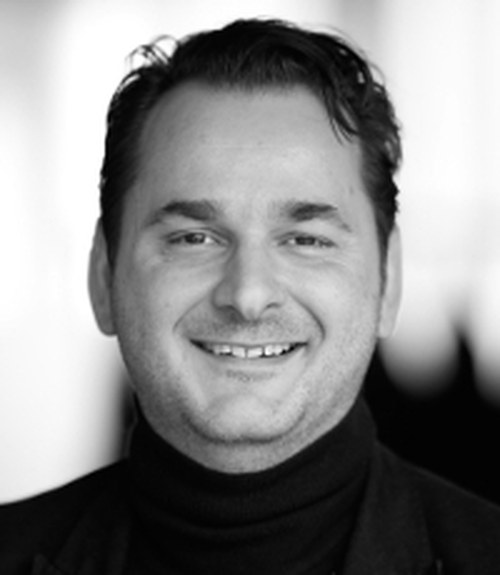
Paolo Gargiulo is an Associate Professor and works at the Medical Technology Center at RU and the University Hospital Landspitali. He studied at TU Wien and finished his PhD in 2008. He has been active in the field of Clinical Engineering, medical image processing and 3-D modeling and tissue engineering. He developed at Landspitali a rapid prototyping service to support surgical planning with over 200 operations planned. He has published 59 papers in peer reviewed international journals and chapters in academic books.
He is consultant of MedEl for the development of larynx pacemaker, co-operating with Össur on the use of EEG to evaluate cortical reorganization in lower limb amputees, with Hjartavernd and NIH to study muscle atrophy or degeneration in aging associated to life styles and co-morbidities, and with Washington University (US) in Brain Modeling project. Since December 2013 Paolo Gargiulo is the director of the Institute of Biomedical and Neural Engineering and the Icelandic Center of Neurophysiology.
Abstract
The seminar will introduce to the ongoing projects and activities taking place at University of Reykjavik within the Institute of Biomedical and Neural Engineering (http://en.ru.is/sse/bne). The main focus will be on qualitative and quantitative assessment of muscles and bone. This seminar will outline methods and applications of threshold-based techniques to assess in vivo muscle and bone tissue distribution in normal and pathological conditions using computed tomography (CT) imaging. These technologies and techniques are used to study bone mechanical proprieties, analyze and quantify muscle morphology, visualize changes with 3D models, develop subject specific numerical profiles, and assess muscle and bone changes during clinical treatments.
Applications of these methodologies are employed: to simulate bone mechanics under particular stressful situation, to depict subject specific muscle profiling associated with age and pathology, to illustrate and quantify muscle degeneration and its partial reversal via Functional Electrical Stimulation (FES), and to highlight recovery following total hip arthroplasty (THA). Furthermore the seminar will summarize some novelties concerning the use of medical imaging and rapid prototyping for surgical planning and prosthetic design.
